How to treat mouth, shell, and scale rot in reptiles.
My appointment book reads that an iguana with “mouth rot” is my 11 o’clock. This rather colorful term is kind of a catch-all phrase for “there is something wrong with my pet’s mouth.” There are other kinds of rots, as well, including “shell rot” and “scale rot.” All are generic terms for problems with the mouth, shell or scales (or skin in general).
These problems are quite common in many herps, and there can be many causes for them. Ulcers, abscesses, abrasions, burns, scabs, parasites, bacterial infections, fungal lesions, viral infections, sloughing areas, tumors, underlying bone abnormalities or necrosis are all medical conditions that may be involved in what is lumped into the term “rot.”
After examining the iguana with the mouth lesion, I determined it actually had a tumor that mimicked the appearance of mouth rot because it involved the bone and soft tissues of the lower mandible. Let’s look into what actually occurs in the conditions called mouth rot, shell rot and scale rot. Identifying an abnormality involving the shell, scales or oral cavity can help herpkeepers decide whether the problem is something they can safely treat, or whether a herp veterinarian’s help is required.
Mouth Rot
Mouth rot is not a specific disease, it is actually a description of a symptom of a disease. It is also called canker or, more correctly, infectious stomatitis. Although infectious stomatitis can occur in any herp, it is most frequently identified in snakes and lizards, and it is not as commonly diagnosed in crocodilians and chelonians (turtles and tortoises).
Most often infectious stomatitis starts off with increased amounts of mucus in the oral cavity and excessive salivation. Oral cavity tissues may have little pinpoint hemorrhages called “petechiae.” These red dots may merge, and the normally pink tissue may change to red or purple. Eventually, if left untreated, the tissue in the area swells, and debris accumulates in the affected areas. Gum tissue may become cracked and may bleed whenever the mouth opens. Tissue may die and cause the damage to spread to deeper tissues, including the bone.
Infection of the bone, called “osteomyelitis,” is very serious and painful, and it requires aggressive treatment. In severe osteomyelitis cases, animals may have loose or missing teeth. Snakes may develop ocular infections if the infection travels into the space between their eyes and spectacles. Pneumonia may result from the chronic weakness and if the reptile inhales organisms into its windpipe and then down into its lungs.
In some cases a normal condition may be misdiagnosed as infectious stomatitis, especially in green iguanas (Iguana iguana). A normal green iguana tongue tip is reddish — not the pink of the rest of the oropharynx — and this may be an erroneous cause for concern.
The causes of infectious stomatitis are many, but the underlying common denominator is usually related to stress, which causes the immune system to function improperly. When this happens, the reptile becomes susceptible to many different disease-causing organisms. These organisms, most frequently bacteria but viruses or fungi in some cases, can begin reproducing unchecked, resulting in serious infection.
In some cases, tumors appear in the tissues surrounding the mouth. If a tumor ulcerates, it may look like infectious stomatitis. For this reason, it is always important for a veterinarian to biopsy the affected tissues in order to procure a diagnosis, especially in cases where ordinary treatment does not resolve the clinical signs.
Herp veterinarians first diagnose infectious stomatitis by taking a thorough history. They will want to know the temperature ranges in the animal’s habitat, the humidity levels, the herp’s actual diet and its behavior in the enclosure. Bacterial culture and sensitivity tests can help to identify what, if any, bacteria are involved. Fungal isolation can identify any fungi involved in the disease process. Gram’s stains of infected areas can show what types of organisms are involved. Cytology can uncover inflammatory cells present in the area. X-rays of the head and perhaps the entire body can also help a vet determine what structures and organ systems are involved, including bones. Blood tests including a complete blood count and plasma chemistry panel can help assess how well a herp is dealing with the problem, and how well its organs are functioning. Biopsy of the affected tissues is often recommended.
Treatment depends on which organisms are involved with the disease and the severity of the infection. Oral cleaning with specific medications may be necessary, but it is important to avoid additional tissue damage by overzealous cleaning of affected tissues. If systemic antibiotics or antifungal agents are needed, either injections or oral medications may be administered.
If the herp is not eating during this time, force-feedings may be required. Only herp vets should perform this activity, or it should be done under their direction. Support care should be provided as needed.
No matter what the treatment, owners must commit to making any necessary husbandry and nutrition changes, or their sick pets won’t respond and heal optimally. If damage is extensive, the tissue may be permanently deformed, which can predispose the herp to repeated events in the future.
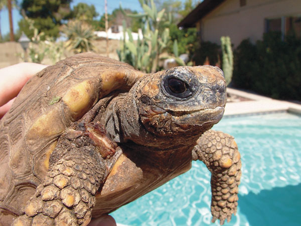
Shell Rot
Shell rot is an all-encompassing name for a group of symptoms in which the shell of turtles and tortoises becomes ulcerated, abscessed or damaged. There are many causes for this condition, and most begin with a scratch or some sort of shell wound. Once that happens, disease-causing bacteria and/or fungi can then enter the wound.
Infection may be mild, affecting just one scute or one area, or widespread. Discoloration or defects may be found in the scutes. Lesions may be pale, or they might show bloody discoloration. Scutes may become loose or fall off entirely in severe cases. Keratin scutes are attached to living bone underneath, so if the infection erodes deeply enough through the shell, it can result in osteomyelitis, a bone infection. Then it is more likely that the infection can spread through the bloodstream to other organs, or the infection may penetrate into the body cavity.
Deep shell abscesses are very serious. Often debris is found in the ulcerated area, which may be foul-smelling. Underneath, the shell may appear moist or bloody. In some cases the abscess may penetrate all the way through the shell and down to the body cavity’s coelomic membrane.
Although infections typically from punctures or bites may occur in terrestrial turtles and tortoises, shell rot infections are most common in aquatic turtles. Because water quality is often involved, dirty water can make things worse very quickly. One form of shell rot found in aquatic turtles is septicemic cutaneous ulcerative disease (SCUD), which may be caused by a variety of bacteria prevalent in tank or pond water. Skin ulcers, decreased appetite and lethargy characterize SCUD, and it may progress to bacteria in the bloodstream, septicemia and eventually death if not treated in time.
Herp veterinarians diagnose shell rot by taking a thorough history, including assessing water quality, habitat, filtration methods, water and air temperature ranges, diet, substrate, number and type of animals in the habitat, lighting, and haulout information. A physical examination identifies where the lesions are located. Then herp vets procure any necessary tests, which may include cytology of the lesions, bacterial culture and sensitivity, fungal isolation, Gram’s staining of lesions, blood tests (including complete blood count and plasma chemistries) and any other recommended tests.
It is very important that chelonians diagnosed with shell rot be kept in a clean environment. A scute scratch or defect can be equated with a human-skin scratch or puncture because the damaged scute can allow bacteria and/or fungi to collect and grow.
Treatment depends on the cause of the infection and the severity of the shell and scute damage. Regardless of the treatment plan, it is vital that the owner commit to making any necessary husbandry or dietary changes. Aquatic turtles are considered the most difficult group of reptiles to properly house and maintain. It is recommended aquatic turtles be fed in a smaller tub or tank easier to clean after mealtime, and then the turtle can be returned to its normal habitat. This helps to keep the permanent habitat clean and free of leftover food and fecal material. Most animals predictably defecate shortly after consuming a meal. If the water temperature has been too cool, submersible aquarium heaters can be safely used, and certain light bulbs can be used to provide heat and ultraviolet light, including UVB, in the turtle’s basking area.
Because a turtle’s shell is attached to underlying bony structures, all ulcers and abscesses are dangerous. Affected chelonians should be “dry docked,” kept dry and warm, except for a short (up to one hour) swim twice daily, so the animal can hydrate and eat. Aquatic turtles won’t eat unless they are in the water. Your herp veterinarian can advise you on the specifics regarding dry docking versus swimming your turtle.
All dead tissue and loose scutes should be removed. In mild cases topical antibiotic ointment may be the only treatment necessary. Ointment should be applied twice daily after the turtle has been removed from the water. Silver-based cream (silver sulfadiazine) is also effective as a topical treatment for many organisms that cause shell rot. Diluted to iced-tea color with warm water, povidone-iodine solution may be used as a flush. Povidone-iodine ointment, applied two to three times per day, may also be painted onto shell lesions for a prolonged contact time. Triple-antibiotic ointment may also be used to cover and treat superficial scute damage. A new topical therapeutic medication called Tricide, which doctors have used to treat antibiotic-resistant bacterial infections in humans, can be used to treat shell rot infections, as well.
Based on bacterial culture and sensitivity results, it may be necessary to administer, usually by injection, systemic antibiotic therapy. The antibiotic chosen should have good ability to penetrate bone if the infection is deep enough to involve bony structures. If surgery is needed to effectively remove the lesions, this should be performed under anesthesia to prevent any pain associated with the procedure. Although pain is a very subjective state, it is often associated with shell rot, so herp vets are likely to administer one of several safe and effective pain medications.
Support care should be administered as needed. Sometimes, intravenous, intracoelomic or subcutaneous fluids may be necessary to treat dehydration and to keep the kidneys functioning. Deep infections may require treatment lasting three to six weeks or longer.
Scale Rot
Scale rot is another all-encompassing term used to describe blister disease, which is vesicular dermatitis or other types of dermatitis in reptiles. Scale rot could also be used to describe bacterial abscesses, burns and the secondary infections that occur after a burn, or infections that occur after skin abrasions.
Many snakes, especially boas and pythons, commonly develop blister disease if they are kept in humidity too high for their species, or if cage substrate remains too damp. Water snakes and garter snakes also have higher chances of developing these diseases in captivity because they often have trouble completely drying off in their environment.
Blisters are small vesicles less than one-half inch in diameter, and bullae are vesicles larger than one-half inch in diameter. Both are thin-walled structures that contain fluid. Vesicles are most often found on the ventral scutes (scales) of snakes from constant contact with moisture.
In most cases the initial vesicles are filled with clear, yellowish fluid and do not contain bacteria. However, if conditions are not corrected, any bacteria found in or on the reptile or its environment may eventually contaminate the vesicles. Mites can also spread bacteria to the vesicles. Rarely some types of fungi, usually opportunistic yeast found in soil, can invade vesicles and result in a skin fungal infection. Once infected with organisms, scales become very red and may begin seeping clear or blood-tinged fluid. Scales may eventually slough off completely.
Sometimes, bacteria may form abscesses at the blister site, and with the next shed ulcers occur once the puslike abscess material is gone. Blisters may coalesce, forming patches of infected skin. Once a superficial infection is treated, skin may shed normally, or it may take a few sheds until the skin heals.
Eventually, if left untreated, bacteria in the blisters can spread through the reptile’s bloodstream and cause septicemia. In large herps this process may take weeks or months, but with small lizards or snakes death can occur in a matter of days.
Immediately place any reptile with suspected scale rot or blister disease in a dry environment with dry substrate, good ventilation, and correct ambient temperature and humidity. Often, placing an infected herp in a clean, dry, healthful environment is all that is necessary to resolve the problem.
Qualified herp veterinarians base a scale rot diagnosis on a thorough history and physical examination. Biopsy of affected scales or scutes may be diagnostic. Other tests may include a Gram’s stain of lesions, bacterial culture and sensitivity, fungal isolation, cytology, blood culture, blood tests (including a complete blood count and plasma chemistry panel) and other applicable tests.
Appropriate antibiotic therapy is critical to proper treatment in all but the most superficial of cases, so it is important that a herp vet sees any herp suspected of suffering from scale rot or a related condition as soon as possible for diagnosis and treatment.
Support care is often necessary in serious cases. Topical treatment with appropriate medications, including silver sulfadiazine, povidone-iodine solution, Tricide or antibiotic creams, may be useful. Infected herps still active and eating have a good chance of successful recovery. Of course, any husbandry or nutritional problems a vet identifies as problematic must be corrected to ensure that this condition does not reoccur.
Prevention Is Key
When it comes to the three rots, prevention is always better than treatment. Ensure that your herp is housed in an appropriate environment that is periodically cleaned and disinfected properly, and offer it a balanced, nutritious diet. Good husbandry practices help prevent herps from ever developing one of these rots. By being a responsible pet owner, you should be able to keep your herp healthy for its entire life.
Potential causes of mouth rot include:
- Improper nutrition. Animals fed an improper diet are much more susceptible to infections.
- Nutritional secondary hyperparathyroidism. This common problem in reptiles may result in a misshapen jaw. Lips are pulled over misshapen bones, and as a result gum tissue develops exposure gingivitis. This syndrome superficially looks like infectious stomatitis, and it will eventually turn into the infection if it is not treated early and properly.
- Mites. Although not a direct cause, mites can contribute to the development of the infection because they can transmit infectious organisms associated with the disease, such as Aeromonas and Pseudomonas bacteria, when they bite and suck blood.
- Trauma. Rostral abrasions from a reptile constantly rubbing its nose on a surface, such as a screen top or glass enclosure, can result in abrasions that allow opportunistic bacteria to enter the tissues and cause infection.
Potential causes of shell rot include:
- Poor water quality. Poor filtration, infrequent water changes or a lack of water conditioners can contribute to shell problems. Water can become fouled when aquatic turtles are fed and live in the same water because uneaten food and fecal material likely will contaminate their habitat.
- Stress and overcrowding.
- Inadequate water temperature.
- Poor nutrition. Animals fed an improper diet are much more susceptible to infections.
- Absence of a haul-out area where turtles can completely dry off.
- Inadequate or poor ultraviolet light.
- Rough cage equipment or bites. In some cases, bites causing shell damage can result in serious damage to the shell. Dog or cat bites can be particularly dangerous because their mouths may contain strains of potentially dangerous bacteria.
Potential causes of scale rot include:
- High humidity.
- Wet substrate.